Pulmonary Embolism | Inferior Vena Cava |
The formation of a blood clot, in a deep leg vein, is known as Deep Vein Thrombosis (DVT) and can be a very serious condition that often causes permanent damage to the leg, known as post-thrombotic syndrome. Early treatment with blood thinners is important to prevent a life-threatening pulmonary embolism but does not treat the existing clot.
Long-term studies show that about 50 percent of people treated with blood thinners alone develop post-thrombotic syndrome. Physicians and patients need to be aware that blood clots cause permanent damage to the leg veins which can lead to serious disability. It is important for DVT patients to be evaluated by an interventional radiologist to determine if catheter-directed thrombolysis is needed to remove the clot. If the clot is not treatable using catheter-directed thrombolysis or the patient is not a candidate, then the interventional radiologist may elect to use an inferior vena cava (IVC) filter.
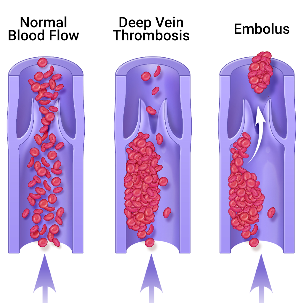
Post-thrombotic syndrome is an under-recognized, but relatively common sequela, or aftereffect, of having DVT if treated with blood thinners (anticoagulation) alone, because the clot remains in the leg. Contrary to popular belief, anticoagulants do not actively dissolve the clot, they just prevent new clots from forming. The body normally dissolves a clot over time, but often the vein becomes damaged in the meantime.
A significant proportion of these patients develop permanent irreversible damage in the affected leg veins and their valves, resulting in abnormal pooling of blood in the leg, chronic leg pain, fatigue, swelling, and in extreme cases, severe skin ulcers. Many patients have to plan their daily activities around their legs, knowing that if they stand or exercise too long, their legs will swell or be painful.
While irreversible damage used to be considered an unusual, long-term sequela, it actually occurs frequently in about 50 percent of people and can develop within two months of developing DVT. Clot removal via interventional catheter-directed thrombolysis in selected cases of DVT can improve quality of life and prevent the debilitating sequela of post-thrombotic syndrome.
Catheter-directed thrombolysis is performed under imaging guidance by interventional radiologists. This procedure, performed in a hospital’s interventional radiology suite, is designed to rapidly break up the clot, restore blood flow within the vein, and potentially preserve valve function to minimize the risk of post-thrombotic syndrome. The interventional radiologist inserts a catheter into the popliteal or other leg vein and threads it into the vein containing the clot using imaging guidance. The catheter tip is placed into the clot and a “clot-busting” drug is infused directly into the thrombus (clot). The fresher the clot, the faster it dissolves—one to two days. Early removal of the blood clots is likely to give patients their best chance to avoid disabling symptoms such as pain, swelling, and ulcer formation in the long run. Any narrowing in the vein that might lead to future clot formation can be identified by venography, an imaging study of the veins, and treated by the interventional radiologist with balloon angioplasty or stent placement.
In patients in whom this is not appropriate and blood thinners are contraindicated, an interventional radiologist can insert a vena cava filter, a small device that functions like a catcher’s mitt to capture blood clots, but allows normal liquid blood to pass. People with symptoms of DVT should first go to an emergency room to seek help, to receive initial treatment with blood thinners to prevent a pulmonary embolism. After treatment with blood thinners, if symptoms such as leg pain and swelling continue, patients should obtain a consultationconsult with an interventional radiologist for further evaluation.
Or schedule an appointment online HERE.
The deep veins that lie near the center of the leg are surrounded by powerful muscles that contract and force deoxygenated blood back to the lungs and heart. One-way valves prevent the back-flow of blood between the contractions. When the circulation of the blood slows down due to illness, injury or inactivity, blood can accumulate or “pool,” which provides an ideal setting for clot formation. One in every 100 people who develops DVT dies. Recently, it has been referred to as “Economy Class Syndrome” due to the occurrence after sitting on long flights.
In the United States alone, 600,000 new cases are diagnosed each year.
Some of these include:
Clinical resolution of pain and swelling and restoration of blood flow in the vein is greater than 85 percent with the catheter-directed technique.
Sitting in one position for a long period of time can increase one’s chances for DVT. To help prevent DVT on long trips:
Left untreated, a deep vein thrombosis can break off and travel in the circulation, getting trapped in the lung, where it blocks the oxygen supply, causing heart failure. This is known as a pulmonary embolism, which can be fatal. With early treatment, people with DVT can reduce their chances of developing a life-threatening pulmonary embolism to less than 1 percent. Blood thinners like heparin and coumadin are effective in preventing further clotting and can prevent a pulmonary embolism from occurring.
Some symptoms of a pulmonary embolism are:
The symptoms are frequently nonspecific and can mimic many other cardiopulmonary events.
In an inferior vena cava filter placement procedure, interventional radiologists use image guidance to place a filter in the inferior vena cava (IVC), the large vein in the abdomen that returns blood from the lower body to the heart.
Blood clots that develop in the veins of the leg or pelvis, a condition called deep vein thrombosis (DVT), occasionally break up and large pieces of the clot can travel to the lungs. An IVC filter traps large clot fragments and prevents them from traveling through the vena cava vein to the heart and lungs, where they could cause severe complications or even death.
Until recently, IVC filters were available only as permanently implanted devices. Newer filters, called optionally retrievable filters, may be left in place permanently or have the option to be removed from the blood vessel later. This removal may be performed when the risk of a clot traveling to the lung has passed. Removal of an IVC filter eliminates any long-term risks of having the filter in place. It does not address the cause of deep vein thrombosis or coagulation. Your referring physician will determine if blood thinners are still necessary.
Inferior vena cava (IVC) filters are placed in patients who have a history of or are at risk of developing blood clots in the legs, including patients:
IVC filters are used when patients cannot be successfully treated by other methods, including blood thinning agents.